top of page
Recapitulating Polyploidization Using Tetraploid iPS Cells
Most cells in our bodies are diploid, containing 23 pairs of chromosomes. They exist as diploid cells because they inherit one copy each of the autosomes (chromosomes 1 through 22) and the 23rd sex chromosome from each parent. However, in certain cell types—such as cardiomyocytes and hepatocytes—a process called “chromosomal duplication” occurs as the cells mature, resulting in an increase in the number of chromosome sets. Approximately 80 percent of adult cardiomyocytes are tetraploid, possessing four copies of each of the 23 chromosome types.
However, in conventional iPS cell-derived cardiomyocytes, this chromosome doubling occurs only to a very limited extent. These cells also remain in an immature, fetal-like state, which has posed a challenge for medical applications.
Therefore, rather than attempting to convert diploid iPS-derived cardiomyocytes into tetraploid cells, we hypothesized that it might be more effective to first convert the iPS cells themselves into tetraploids and then differentiate them into cardiomyocytes. Indeed, we successfully established human tetraploid iPS cells through cell fusion (Fig. 1) and achieved their differentiation into cardiomyocytes. As expected, cardiomyocytes derived from tetraploid iPS cells exhibited more mature characteristics, such as strong and fast contractions, thereby expanding the potential for medical applications using iPS cell-derived cardiomyocytes (Fig. 2, Nakajima, Commun Biol 2026).
It is known that chromosome doubling occurs not only in cardiomyocytes but also in several other cell types, including hepatocytes, skeletal muscle cells, and osteoclasts. Furthermore, there are reports indicating that chromosome doubling in cells such as neurons—which are normally diploid—is a sign of aging or disease. We aim to recapitulate the chromosome doubling that occurs in these cells using tetraploid iPS cells, utilize them to generate more mature cells and as models of aging and disease, and ultimately develop treatments for these conditions.
Fig. 1
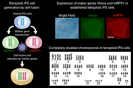
Fig. 2

One-bp Substitution in the 3 Billion-bp Human Genome
Genome editing allows for any manipulations of the genome in human iPS cells, which can be applied for disease modeling and cell transplantation therapy. However, it had been difficult to substitute only one base-pair (bp) in the 3 billion-bp human genome without leaving artificial DNA sequences. One-bp substitution by genome editing can serve as a versatile platform for various applications, because many diseases are caused by single-bp mutations.
We developed a method for isolation of iPS cells with only single-bp substitutions by combining CRISPR-Cas9, TALEN, and digital PCR (Fig. 3, Miyaoka Nature Methods 2014).
By using our method, we can introduce single-bp substitutions at any locations in the genome (Fig. 4).

➡︎
➡︎
⬅︎
⬅︎
Fig. 3
Fig. 4

Modeling Disease in a Dish to Study Pathogenesis
Fig. 5

By editing the genome in iPS cells, we can study pathogenesis of genetic disorders in any cell types in a dish. For example, a point mutation of cardiomyopathy introduced into iPS cells caused abnormal sarcomere structure (a functional unit of muscle contraction visualized as red stripes), when these cells were differentiated into cardiomyocytes (Fig. 5). We revealed a pathogenic mechanism by abnormal aggregation of RNA and proteins unexpectedly shared between cardiomyopathy and neurodegenerative diseases.
Wild-type cell
Mutant cell
ACTININ/Nucleus
Seeking for Precise Genome Editing Conditions for Therapy
iPS cells from patients with genetic disorders maintain the causative mutations. However, if we correct these mutations, the patient-derived iPS cells can potentially be used for cell therapy. For this purpose, we need genome editing that does not cause any unwanted damages to the genome. We will correct pathogenic mutations such as ones of Wilson's disease (a genetic liver disorder) in iPS cells with diverse genome editing conditions to evaluate their accuracy by using highly sensitive digital PCR and other techniques.
bottom of page